Wednesday, 19 September 2007

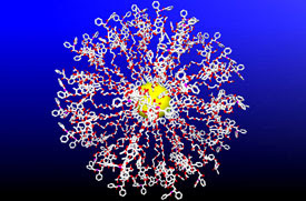
Taxol Bristle Ball

Cancer-clogging drugs loaded onto nanospheres from Rice University

Rice chemists have discovered a way to load dozens of molecules of the anticancer drug paclitaxel onto tiny gold spheres. The result is a ball many times smaller than a living cell that literally bristles with the drug.

Paclitaxel, which is sold under the brand name Taxol®, prevents cancer cells from dividing by jamming their inner works.

First isolated from the bark of the yew tree in 1967, paclitaxel is one of the most widely prescribed chemotherapy drugs in use today. The drug is used to treat breast, ovarian and other cancers. Paclitaxel works by attaching itself to structural supports called microtubules, which form the framework inside living cells. To divide, cells must break down their internal framework, and paclitaxel stops this process by locking the support into place.

Since cancer cells divide more rapidly than healthy cells, paclitaxel is very effective at slowing the growth of tumors in some patients. However, one problem with using paclitaxel as a general inhibitor of cell division is that it works on all cells, including healthy cells that tend to divide rapidly. This is why patients undergoing chemotherapy sometimes suffer side effects like hair loss and suppressed immune function.

"Ideally, we'd like to deliver more of the drug directly to the cancer cells and reduce the side effects of chemotherapy," Zubarev said. "In addition, we'd like to improve the effectiveness of the drug, perhaps by increasing its ability to stay bound to microtubules within the cell."

The new delivery system centers on a tiny ball of gold that's barely wider than a strand of DNA. Finding a chemical process to attach a uniform number of paclitaxel molecules to the ball - without chemically altering the drug - was not easy. Only a specific region of the drug binds with microtubules. This region of the drug fits neatly into the cell's support structure, like a chemical "key" fitting into a lock. Zubarev and Gibson knew they had to find a way to make sure the drug's key was located on the face of each bristle.

Zubarev and Gibson first designed a chemical "wrapper" to shroud the key, protecting it from the chemical reactions they needed to perform to create the ball. Using the wrapped version of the drug, they undertook a series of reactions to attach the drug to linker molecules that were, in turn, attached to the ball. In the final step of the reaction, they dissolved the wrapper, restoring the key."We are already working on follow-up studies to determine the potency of the paclitaxel-loaded nanoparticles," Zubarev said. "Since each ball is loaded with a uniform number of drug molecules, we expect it will be relatively easy to compare the effectiveness of the nanoparticles with the effectiveness of generally administered paclitaxel."
_________________________________________________________
_________________________________________________________

Saturday, 1 September 2007

New Cancer Weapon


Nuclear Nanocapsules

Rice University chemists have found a way to package some of nature's most powerful radioactive particles inside DNA-sized tubes of pure carbon - a method they hope to use to target tiny tumors and even lone leukemia cells.


"There are no FDA-approved cancer therapies that employ alpha-particle radiation," said lead researcher Lon Wilson, professor of chemistry. "Approved therapies that use beta particles are not well-suited for treating cancer at the single-cell level because it takes thousands of beta particles to kill a lone cell.

By contrast, cancer cells can be destroyed with just one direct hit from an alpha particle on a cell nucleus."

In the study, Wilson, Rice graduate student Keith Hartman, University of Washington (UW) radiation oncologist Scott Wilbur and UW research scientist Donald Hamlin, developed and tested a process to load astatine atoms inside short sections of carbon nanotubes.

Because astatine is the rarest naturally occurring element on Earth - with less than a teaspoon estimated to exist in the Earth's crust at any given time - the research was conducted using astatine created in a UW cyclotron.

Astatine, like radium and uranium, emits alpha particles via radioactive decay. Alpha particles, which contain two protons and two neutrons, are the most massive particles emitted as radiation. About 4,000 times more massive than the electrons emitted by beta decay - the type of radiation most commonly used to treat cancer.

"It's something like the difference between a cannon shell and a BB," Wilson said. "The extra mass increases the amount of damage alpha particles can inflict on cancer cells."

The speed of radioactive particles is also an important factor in medical use. Beta particles travel very fast. This, combined with their small size, gives them significant penetrating power.

In cancer treatment, for example, beams of beta particles can be created outside the patient's body and directed at tumors. Alpha particles move much more slowly, and because they are also massive, they have very little penetrating power. They can be stopped by something as flimsy as tissue paper.

"The unique combination of low penetrating power and large particle mass make alpha particle ideal for targeting cancer at the single-cell level," Wilson said. "The difficulty in developing ways to use them to treat cancer has come in finding ways to deliver them quickly and directly to the cancer site."

In prior work, Wilson and colleagues developed techniques to attach antibodies to carbon fullerenes like nanotubes. Antibodies are proteins produced by white blood cells. Each antibody is designed to recognize and bind only with a specific antigen, and doctors have identified a host of cancer-specific antibodies that can be used to kill cancer cells.

In follow-up research, Wilson hopes to test the single-celled cancer targeting approach by attaching cancer-specific antibodies to astatine-loaded nanotubes.

One complicating factor in any astatine-based cancer therapy will be the element's short, 7.5-hour half-life. In radioactive decay, the term half-life refers to the time required for any quantity of a substance to decay by half its initial mass.

Due to astatine's brief half-life, any treatment must be delivered in a timely way, before the particles lose their potency.
________________________________________________________
________________________________________________________

Friday, 24 August 2007

Area responsible for Self Control

The area of the brain responsible for self-control is separate from the area associated with taking action.

Image Above: Brain area in the fronto-median cortex that was activated when participants intentionally withhold a planned action in the last moment

The results illuminate a very important aspect of the brain's control of behavior, the ability to hold off doing something after you've developed the intention to do it-one might call it 'free won't' as opposed to free will," says Martha Farah, PhD, of the University of Pennsylvania. "It is very important to identify the circuits that enable 'free won't' because of the many psychiatric disorders for which self-control problems figure prominently-from attention deficit disorder to substance dependence and various personality disorders." Farah was not involved in the experiment.

The findings broaden understanding of the neural basis for decision making, or free will, and may help explain why some individuals are impulsive while others are reluctant to act, says lead author Marcel Brass, PhD, of the Max Planck Institute for Human Cognitive and Brain Sciences and Ghent University. Brass and Patrick Haggard, PhD, of University College London, used functional magnetic resonance imaging (fMRI) to study the brain activity of participants pressing a button at times they chose themselves. They compared data from these trials to results when the participants prepared to hit the button, then decided to hold back or veto the action.

Fifteen right-handed participants were asked to press a button on a keyboard. They were asked to choose some cases in which they stopped just before pressing the button. Participants also indicated on a clock the time at which they intended to press the button or decided to hold back. When Brass and Haggard compared fMRI images of the two scenarios, they found that pulling back yielded activity in the dorsal fronto-median cortex (dFMC), an area on the midline of the brain directly above the eyes, which did not show up when participants followed through and made the action. In addition, those who chose to stop the intended action most often showed greatest contrast in dFMC activity.

"The capacity to withhold an action that we have prepared but reconsidered is an important distinction between intelligent and impulsive behavior," says Brass, "and also between humans and other animals."

Future study will involve methods with a better time resolution such as EEG to determine whether the inhibitory process could operate in the brief time period between the time of conscious intention and the point of no return for motor output.

Image: Max Planck Institute for Human Cognitive and Brain Sciences
________________________________________________________
________________________________________________________
Milestone In The Regeneration Of Brain Cells
Brain Cells Work Differently Than Previously Thought
One Step Closer To Transplanting Stem Cells In The Brain
Researchers Identify Brain Network That May Help Prevent Or Slow Alzheimer's
________________________________________________________
________________________________________________________

Tuesday, 21 August 2007

Ancestors help Bird Memory


Birds learn to fly with a little help from their Ancestors

A University of Sheffield researcher has discovered that the reason birds learn to fly so easily is because latent memories may have been left behind by their ancestors.

It is widely known that birds learn to fly through practice, gradually refining their innate ability into a finely tuned skill, and these skills may be easy to refine because of a genetically specified latent memory for flying.

Dr Dr Jim Stone from the University of Sheffield´s Department of Psychology, used simple models of brains called artificial neural networks and computer simulations to test his theory.

He discovered that learning in previous generations indirectly induces the formation of a latent memory in the current generation and therefore decreases the amount of learning required. These effects are especially pronounced if there is a large biological 'fitness cost' to learning, where biological fitness is measured in terms of the number of offspring each individual has.

The beneficial effects of learning also depend on the unusual form of information storage in neural networks. Unlike computers, which store each item of information in a specific location in the computer's memory chip, neural networks store each item distributed over many neuronal connections. If information is stored in this way then evolution is accelerated, explaining how complex motor skills, such as nest building and hunting skills, are acquired by a combination of innate ability and learning over many generations.

However not every bird automatically knows how to fly.
A crowling which has been blown from its nest, had a great fall and hurt its legs, may never fly - though there be nothing wrong with its wings at all. Birds learn fear, pain and doubt too. - Q9

________________________________________________________
________________________________________________________
New Caledonian Crows Find Two Tools Better Than One
Uncertainties Of Savanna Habitat Drive Birds To Cooperative Breeding
Birds With Child-care Assistance Invest Less In Eggs @ Science Daily
Are Artist's Born or Taught from Hank @ Scientific Blogging
________________________________________________________
________________________________________________________

Friday, 10 August 2007

Functioning Neuron Produced


Scientists with the Institute of Stem Cell Biology and Medicine at UCLA were able to produce from human embryonic stem cells a highly pure, large quantity of functioning neurons that will allow them to create models of and study diseases such as Alzheimer's, Parkinson's, prefrontal dementia and schizophrenia.

Researchers previously had been able to produce neurons - the impulse-conducting cells in the brain and spinal cord - from human embryonic stem cells. However, the percentage of neurons in the cell culture was not high and the neurons were difficult to isolate from the other cells.

UCLA's Yi Sun, an associate professor of biobehavioural sciences, and Howard Hughes Medical Institute investigator Thomas Südhof at the University of Texas Southwestern Medical Center were able to produce 70 to 80 percent of neurons in cell culture. Sun and Südhof also were able to isolate the neurons and determine that they had a functional synaptic network, which the neurons use to communicate. Because they were functional, the neurons can be used to create a variety of human neurological disease models.

"Previously, the system to grow and isolate neurons was very messy and it was unknown whether those neurons were functioning," Sun said. "We're excited because we have been able to purify so many more neurons out of the cell culture and they were, surprisingly, healthy enough to form synapses. These cells will be excellent for doing gene expression studies and biochemical and protein analyses."

Sun's method prodded human embryonic stem cells to differentiate into neural stem cells, the cells that give rise to neurons. When the time was right, Sun's team added protein growth factors into the cell culture that stopped the neural stem cells from self-renewing and prodded them into differentiating into neurons.

To isolate the cells, Sun and her team added an enzyme that digests a sort of protein matrix that holds cells in culture together. The neurons could then be separated from the neural stem cells that had not yet differentiated, a sort of chemical round-up that isolated the neurons. The cells were then put into a cell strainer that allowed passage through of the isolated neurons.

The large number of pure neurons produced will allow Sun and her team to study their biological form and structure, the genes they express, the development of synapses and the electric and chemical communication activities within the synapse network.

"We will be able to study the cellular properties of neurons in a very defined way that will maybe tell us what goes wrong in diseases such as Alzheimer's and Parkinson's," Sun said. "We're currently creating many models of human neurological diseases that may provide the answers we're looking for. We don't know what causes prefrontal dementia, Huntington's disease or schizophrenia. The key is likely in the quality of neuronal communications. By studying the chemical and electrical transmissions, we may be able to determine what goes wrong that leads to these debilitating diseases and find a way to stop or treat it."

Sun will be among the first researchers to be able to study true neuron function.

A second important discovery in Sun's study showed that two embryonic stem cells lines derived in similar manners, and therefore expected to behave similarly when differentiating, did not. Using the same techniques to prod the two embryonic stem cells lines to differentiate, Sun found that one line had a bias to become neurons that are found in the forebrain. The other line differentiated into neurons found in rear portions of the brain and spinal cord. The finding was surprising, and significant, Sun said.

"The realization that not all human embryonic stem cell lines are born equal is critical," Sun said. "If you're studying a disease found in a certain part of the brain, you should use a human embryonic stem cell line that produces the neurons from that region of the brain to get the most accurate results from your study.

Huntington's disease, for example, is a forebrain disease, so the neurons should be differentiated from a cell line that is biased to produce neurons from the forebrain."Sun said there are ways to prod an embryonic stem cell line biased to become neurons found in the rear brain to become neurons found in the forebrain. However, there are limits to how much prodding can be done.

Sun and her team confirmed that the two embryonic stem cell lines were different through gene expression analysis - neurons that perform different functions in different parts of the brain express different genes.

The cell line prone to becoming neurons found in the forebrain expressed genes typically found in those neurons, while the other line expressed genes found in the rear brain and spinal cord.

The team now are studying why the two human embryonic stem cell lines have biases to become different types of neurons. "If we knew that, we might be able to tweak or alter whatever is driving the bias so that limitation in the stem cell line could be bypassed," Sun said.

Study results were recently published in an early online edition of the journal Proceedings of the National Academy of Sciences.

Functioning Neurons From Human Embryonic Stem Cells Produced
UCLA Institute of Stem Cell Biology
________________________________________________________
________________________________________________________

Wednesday, 8 August 2007

Pain Perception

Treatment of pain-related suffering requires knowledge of how pain signals are initially interpreted and subsequently transmitted and perpetuated.

Clinical pain is a serious public health issue.
Our understanding of the neural correlates of pain perception in humans has improved with the advent of neuroimaging. Relating neural activity changes to the varied pain experiences has led to an increased awareness of how factors (e.g., cognition, emotion, context, injury) can separately influence pain perception.

It has been suggested that the brainstem plays a pivotal role in gating the degree of nociceptive transmission so that the resultant pain experienced is appropriate for the particular situation of the individual.

Pain that persists for more than three months is defined as chronic and as such is one of largest medical health problems in the developed world. While the management and treatment of acute pain is reasonably good, the needs of chronic pain sufferers are largely unmet, creating an enormous emotional and financial burden to sufferers, carers, and society.

The mechanisms that contribute to the generation and maintenance of a chronic pain state are increasingly investigated and better understood. A consequent shift in mindset that treats chronic pain as a disease rather than a symptom is accelerating advances in this field considerably.

Pain is a conscious experience, an interpretation of the nociceptive input influenced by memories, emotional, pathological, genetic, and cognitive factors. Resultant pain is not necessarily related linearly to the nociceptive drive or input; neither is it solely for vital protective functions. This is especially true in the chronic pain state. Furthermore, the behavioral response by a subject to a painful event is modified according to what is appropriate or possible in any particular situation. Pain is, therefore, a highly subjective experience “an unpleasant sensory and emotional experience associated with actual or potential tissue damage, or described in terms of such damage.”

By its very nature, pain is therefore difficult to assess, investigate, manage, and treat. Figure 1 (above) illustrates the mixture of factors that we know influence nociceptive inputs to amplify, attenuate, and color the pain experience.

Because pain is a complex, multifactorial subjective experience, a large distributed brain network is subsequently accessed during nociceptive processingm and was first described this as the pain “neuromatrix,” but it's now more commonly referred to as the “pain matrix”; simplistically it can be thought of as having lateral (sensory-discriminatory) and medial (affective-cognitive-evaluative) neuroanatomical components. However, because different brain regions play a more or less active role depending upon the precise interplay of the factors involved in influencing pain perception (e.g., cognition, mood, injury, and so forth), what comprises the pain matrix is not unequivocally defined.

For both chronic and acute pain sufferers, mood and emotional state has a significant impact on the resultant pain perception and ability to cope. For example, it is a common clinical and experimental observation that anticipating and being anxious about pain can exacerbate the pain experienced. Anticipating pain is highly adaptive; we all learn in early life to avoid hot pans on stoves and not to put your finger into a candle flame. However, for the chronic pain patient it becomes maladaptive and can lead to fear of movement, avoidance, anxiety, and so forth.

Another negative cognitive and mood affect that impacts pain is catastrophizing. This construct incorporates magnification of pain-related symptoms, rumination about pain, feelings of helplessness, and pessimism about pain-related outcomes, and it is defined as a set of negative emotional and cognitive processes. A study on fibromyalgia patients found that pain catastrophizing, independent of the influence of depression, was significantly associated with increased activity in brain areas related to anticipation of pain (medial frontal cortex, cerebellum), attention to pain (dorsal ACC, dorsolateral prefrontal cortex), emotional aspects of pain (claustrum, closely connected to amygdala), and motor control.

Clearly, these results support the notion that catastrophizing influences pain perception through altering attention and anticipation, as well as heightening emotional responses to pain.

It is interesting to speculate whether activity in such “emotional” brain regions due to chronic pain impacts performance in tasks requiring emotional decision making. A card game developed to study emotional decision making, chronic pain patients displayed a specific cognitive deficit compared to controls, suggesting such an impact might exist in everyday life.

Such experiments are hard to reproduce in animal studies.

As the problem of pain and the key role of the brain becomes increasingly well recognized, more research is being directed toward a better understanding of the underlying mechanisms. Some of the newest and more novel areas of investigation are briefly summarized here.

The recent finding that significant atrophy exists in the brains of chronic pain patients highlights the need to perform more advanced structural imaging measures and image analyses to quantify fully these effects.

Determining what the possible causal factors are that produce such neurodegeneration is difficult. Candidates include the chronic pain condition itself (i.e., excitotoxic events due to barrage of nociceptive inputs), the pharmacological agents prescribed, or perhaps the physical lifestyle change subsequent to becoming a chronic pain patient.
_________________________________________________________
Backache Sufferers Who Fear Pain Change Movements from Science Daily
_________________________________________________________
_________________________________________________________
Monday, 6 August 2007

Music on the Brain


Music moves brain to pay attention

Using brain images of people listening to short symphonies by an obscure 18th-century composer, a research team from the Stanford University School of Medicine has gained valuable insight into how the brain sorts out the chaotic world around it.

The research team showed that music engages the areas of the brain involved with paying attention, making predictions and updating the event in memory. Peak brain activity occurred during a short period of silence between musical movements—when seemingly nothing was happening.

Beyond understanding the process of listening to music, their work has far-reaching implications for how human brains sort out events in general. The findings are published in the Aug. 2 issue of Neuron.

The researchers caught glimpses of the brain in action using functional magnetic resonance imaging, or fMRI, which gives a dynamic image showing which parts of the brain are working during a given activity. The goal of the study was to look at how the brain sorts out events, but the research also revealed that musical techniques used by composers 200 years ago help the brain organize incoming information.

The team used music to help study the brain’s attempt to make sense of the continual flow of information the real world generates, a process called event segmentation. The brain partitions information into meaningful chunks by extracting information about beginnings, endings and the boundaries between events.

See Video & Read more @ Stanford University School Of Medicine news release
________________________________________________________
________________________________________________________
Nerve Cell Modulator Could Help With Mood Disorders
Virtual Navigation - Rethinking How Columbus Mentally Found His Way
Connectivity Between Brain Areas Leads To Human Knowledge
Prefrontal Cortex Differences Mean Richer Memories For Adults
________________________________________________________
________________________________________________________

Thursday, 2 August 2007

Neuro Genesis in Korea?

Toward An Alternative To Stem Cells For Treating Chronic Brain Diseases
Image Courtesy of Injae Shin, Yonsei University, Korea.

Scientists in Korea are reporting the first successful use of a drug-like molecule to transform human muscle cells into nerve cells. This advance could lead to new treatments for stroke, Alzheimer's disease, Parkinson's disease and other neurological disorders.

The researchers exposed immature mouse muscle cells myoblasts (image left) growing in laboratory cell cultures to neurodazine, a synthetic small molecule. After one week, 40-50 percent of the myoblasts were transformed into cells that resembled both the structure and function of nerve cells (image right), including expression of neuron-specific proteins. Additional studies showed a similar transformation in a group of human skeletal muscle cells that were exposed to the same chemical for several days, they add.

"In conclusion, we have developed the first small molecule that can induce neurogenesis of non-pluripotent myoblasts and the cells derived from mature, human skeletal muscle," the report states. "These studies build upon recent research illustrating the value of chemical approaches for providing tools that differentiate lineage-committed cells into other cell types."
________________________________________________________
MS breakthrough new genetic risk factors discovered
Rare Example of Darwinism Seen in Action - UC Riverside release
Factor That Causes Embryonic Stem Cell Specialization from Scientific Blogging
________________________________________________________
________________________________________________________

Friday, 27 July 2007

How Real is Synesthesia?


Hearing Colours And Seeing Sounds:
How Real Is Synesthesia?


In the psychological phenomenon known as "synesthesia," individuals' sensory systems are a bit more intertwined than usual. Some people, for example, report seeing colours when musical notes are played.

One of the most common forms is grapheme-colour synesthesia, in which letters or numbers (collectively called "graphemes") are highlighted with particular colours. Although synesthesia has been well documented, it is unknown whether these experiences, reported as vivid and realistic, are actually being perceived or if they are a byproduct of some other psychological mechanism such as memory.

New research published in the June issue of Psychological Science, a journal of the Association for Psychological Science, sheds some light on the veracity of these perceptions.

When anyone views a particular colour, specific neurons in the visual cortex area of our brain are activated. These specific neurons will deactivate, however, if a colour from the opposite end of the spectrum is presented.

Any neuron activated when the colour blue is present will deactivate when it's exact opposite, yellow, comes into the visual field.

Read more from Science Daily
_______________________________________________________
_______________________________________________________
UC Irvine scientists unveil the 'face' of a new memory Breakthrough study also links learning to a specific chemical process in brain cells
_______________________________________________________
_______________________________________________________

Thursday, 19 July 2007

Protein's Internal Motion


Researchers at the University of Pennsylvania School of Medicine are the first to observe and measure the internal motion inside proteins, or its “dark energy.” This research, appearing in the current issue of Nature, has revealed how the internal motion of proteins affects their function and overturns the standard view of protein structure-function relationships, suggesting why rational drug design has been so difficult.

“The situation is akin to the discussion in astrophysics in which theoreticians predict that there is dark matter, or energy, that no one has yet seen,” says senior author A. Joshua Wand, PhD, Benjamin Rush Professor of Biochemistry. “Biological theoreticians have been kicking around the idea that proteins have energy represented by internal motion, but no one can see it. We figured out how to see it and have begun to quantify the so-called ‘dark energy’ of proteins.”

Proteins are malleable in shape and internal structure, which enables them to twist and turn to bind with other proteins. “The motions that we are looking at are very small, but very fast, on the time scale of billions of movements per second,” explains Wand. “Proteins just twitch and shake.” The internal motion represents a type of energy called entropy.

Current models of protein structure and function used in research and drug design often do not account for their non-static nature. “The traditional model is almost a composite of all the different conformations a protein could take,” says Wand.

The researchers measured a protein called calmodulin and its interactions with six other proteins when bound to a protein partner one at a time. These binding partners included proteins important in smooth muscle contraction and a variety of brain functions.

Using nuclear magnetic resonance spectroscopy, the investigators were able to look at the changes in the internal motion of calmodulin itself in each of the six different protein binding situations. They found a direct correlation between a change in calmodulin’s entropy – a component of its stored energy – and the total entropy change leading to the formation of the calmodulin-protein complex.


Finding out the contribution from individual proteins versus the entropy, or movement, of the entire protein complex has been more difficult and has been overcome in this study. From this individual contribution they deduced that changes in the entropy of the protein are indeed important to the process of calmodulin binding its partners.

“Before these unexpected results, most researchers in our field would have predicted that entropy’s contribution to protein-protein interactions would be zero or negligible,” says Wand. “But now it’s clearly an important component of the total energy in protein binding.”

Because of this new information, the researchers suggest that the entropy component may explain why drug design fails more often than it works. Currently, drugs are designed generally based on the precise structures of their biological targets, active regions on proteins that are intended to inhibit key molecules. However, the number of designed molecules actually binding to their targets is low for many engineered molecules. “We think that this is because the design is based on a model of a static protein, not the moving, hyper protein that is constantly changing shape,” say Wand. “We need to figure out how this new information fits in and perhaps drug design could be significantly improved.”

Future directions include understanding whether the principles revealed by this study are universal and impact the thousands of protein-protein interactions that underlie biology and disease. As Wand explains, “Protein-protein interactions are central to ‘signalling’, which is often the molecular origin of diseases. Cancer, diabetes, and asthma are three important examples. We are currently looking at the role of protein entropy in the control of critical signaling events in all three.”

Artist rendering of calmodulin molecule depicting protein "dark energy."
Image Credit: Mary Leonard and Michael Marlow, UP School of Medicine.

Proteins' Internal Motion, Implications For Drug Design Science Daily Releases
New proteomics research promises to revolutionize biomedical discovery
Protein Pulling: Learning How Proteins Fold By Pulling Them Apart
_________________________________________________________
_________________________________________________________

Tuesday, 17 July 2007

Unravelling the Physics of DNA


Researchers at Duke University's School of Engineering have uncovered a missing link in scientists' understanding of the physical forces that give DNA its famous double helix shape.

The stability of DNA is fundamental to life. To create accurate models of DNA to study its interaction with proteins or drugs, you need to understand the basic physics of the molecule. For that, you need solid measurements of the forces that stabilize DNA.

Each DNA strand includes a sugar and phosphate "backbone" attached to one of four bases, which encode genetic sequences. The strength of the interactions within individual strands comes largely from the chemical attraction between the stacked bases. But the integrity of double-stranded DNA depends on both the stacking forces between base units along the length of the double helix and on the pairing forces between complementary bases, which form the rungs of the twisted ladder.

Earlier studies have focused more attention on the chemical bonds between opposing bases, measuring their strength by "unzipping" the molecules' two strands. Studies of intact DNA make it difficult for researchers to separate the stacking from the pairing forces.

To get around that problem in the new study, the Duke team used an atomic force microscope (AFM) to capture the "mechanical fingerprint" of the attraction between bases within DNA strands. The bonds within the molecules' sugar and phosphate backbones remained intact and therefore had only a minor influence on the force measurements.

Researchers tugged on individual strands that were tethered at one end to gold and measured the changes in force as they pulled. The AFM technique allows precise measurements of forces within individual molecules down to one pico-Newton--a trillionth of a Newton. For a sense of scale, the force of gravity on a two-liter bottle of soda is about 20 Newtons.

They captured the range of stacking forces by measuring two types of synthetic DNA strands: some made up only of the base thymine, which is known to have the weakest attraction between stacked units, and some made up only of the base adenine, known to have the strongest stacking forces. Because of those differences in chemical forces, the two types of single-stranded DNA take on different structures. Single strands of adenine coil in a fairly regular fashion to form a helix of their own, while thymine chains take on a more random shape.

The pure adenine strands exhibited an even more complex form of elasticity than had been anticipated, the researchers reported. As they stretched the adenine chains with increasing force, the researchers noted two places--at 23 and 113 pico-Newtons--where their measurements leveled off.

"Those plateaus reflect the breaking and unfolding of the helix," professor of mechanical engineering and materials sciences at Duke, Marszalek explained. With no bonds between bases to break, the thymine chains' showed little resistance to extension and no plateau.

Based on the known structure of the single stranded DNA molecules, they had expected to see only one such plateau as the stacking forces severed. Exactly what happens at the molecular level at each of the two plateaus will be the subject of continued investigation.
_________________________________________________________
_________________________________________________________

Friday, 13 July 2007

White Blood Cells Are Picky About Sugar


Biology textbooks are blunt--neutrophils are mindless killers. These white blood cells patrol the body and guard against infection by bacteria and fungi, identifying and destroying any invaders that cross their path.

But new evidence, which may lead to better drugs to fight deadly pathogens, indicates that neutrophils might actually distinguish among their targets.

A scientist in the lab of Whitehead Member Gerald Fink has discovered that neutrophils recognize and respond to a specific form of sugar called beta-1,6-glucan on the surface of fungi. This sugar comprises just a small fraction of the fungal cell wall, much less than another sugar with a slightly different chemical conformation called beta-1,3-glucan. Because the scarce form of the sugar elicits a much stronger reaction from immune cells than the abundant one, it appears that neutrophils can distinguish between two nearly identical chemicals.

"These results show that engulfment and killing by neutrophils varies, depending on cell wall properties of the microbe," explains Whitehead postdoctoral researcher Ifat Rubin-Bejerano, first author on the paper, which appears July 11 in the journal Cell Host & Microbe. "We showed that neutrophils respond in a completely different way to slight changes in sugar composition. If we are able to use this unique sugar to excite the immune system, it may help the human body fight infection."

"Previously, everyone thought that these key cells of the immune system weren't picky and would eat anything that looked foreign," adds Fink, who is also an MIT professor of biology. "Ifat's work has shown that the cells aren't little Pac-Men, but can discriminate one pathogen from another."

Rubin-Bejerano had evidence that neutrophils respond to beta-glucan. After coating tiny beads with a variety of substances (including beta-1,3-glucan and beta-1,6-glucan), she exposed them to the neutrophils and was surprised to see a striking difference in their response to the two sugars. The neutrophils quickly engulfed many of the beads coated with beta-1,6-glucan, but only a few of those covered in beta-1,3-glucan.

Previous studies indicated that blood serum (basically blood minus cells) helps neutrophils recognize their enemies, so Rubin-Bejerano decided to look for clues to their response in this mixture. She identified several proteins in serum that bind to beta-1,6-glucan, but not beta-1,3-glucan, and then pinpointed a molecule on the surface of the neutrophil that recognizes these proteins.

To link her experiments back to real fungi, Rubin-Bejerano worked with the pathogen Candida albicans, which is the most common fungus in blood stream infections. She used an enzyme to digest beta-1,6-glucan from the fungal cell wall, leaving the beta-1,3-glucan intact. She then unleashed the neutrophils on these altered cells and observed a 50 percent reduction in the immune response.

Our bodies maintain a fine balance between the immune system and microbes. Antibiotics and antifungals tilt the balance in favor of the immune system by targeting the microbes directly. A substance like beta-1,6-glucan could help tilt this balance further by stimulating immune cells.

Rubin-Bejerano's work offers hope for combating the growing problem of microbial infections, which can seriously threaten human health--particularly in patients with compromised immune systems. In fact, Rubin-Bejerano co-founded a company called ImmuneXcite to explore this possibility.

Source: Whitehead Institute for Biomedical Research
_________________________________________________________
_________________________________________________________
Has Science Unearthed The Holy Grail Of Pain Relief?
New Way To Target And Kill Antibiotic-resistant Bacteria Found
Human Embryonic Stem Cells Are The Ultimate Perpetual Fuel Cell, Study Shows
_________________________________________________________
_________________________________________________________

Monday, 9 July 2007

How DNA strands separate


Cornell researchers have answered a fundamental question about how two strands of DNA, known as a double helix, separate to start a process called replication, in which genes copy themselves.

Credit: Chris Pelkie and Daniel Ripoll/Cornell Theory Center


This image shows a DNA double helix (green and purple strands) being separated by a helicase enzyme (green globule) at the junction where the two strands fork. To show that helicases actively separate the two strands of DNA, the researchers attached one end of a DNA strand to a microscope cover slip and attached the end of the other DNA strand to a micron-sized plastic bead. The bead was then trapped in a tightly focused laser beam (red), which allowed the researchers to measure the motion of the helicase as it unwound the DNA.

The research, published in the current issue of the journal Cell, examined the role of an enzyme called a helicase, which plays a major role in separating DNA strands so that replication of a single strand can occur.

Scientists have known that helicases bind to the area of a double helix where the two strands fork away from each other, like the free ends of two pieces of thread wound around each other. The forked area opens and closes very rapidly. But scientists have debated whether helicases actively separate the two strands at the fork or if they passively wait for the fork to widen on its own.

The research found that the helicase appears to actively exert a force onto the fork and separate the two strands.

"A simple passive unwinding mechanism does not explain our data," said Michelle Wang, associate professor of physics and the paper's senior author.

"Defects in helicases are associated with many human diseases, ranging from predisposition to cancer to premature aging," said co-author Smita Patel, a biochemistry professor at the Robert Wood Johnson Medical School in Piscataway, N.J. "Helicases are involved in practically all DNA and RNA metabolic processes."

The researchers made their discovery by anchoring one end of one of the strands in a double helix to the surface of a microscope cover slip. The end of the other strand was attached to a micron-sized plastic bead. They then focused a laser beam on the tiny bead and trapped the bead in place within the beam of light. This setup allowed the researchers to measure the position and force on the bead, creating a very precise sensor of the helicase motion. As the helicase moved toward the fork and the double helix unwound, the tension on the two strands lessened. Using statistical mechanics models, the researchers could then compare actual measurements of movement with predictions based on both active and passive scenarios.

"The unwinding has to have some active component to it, and based on our data, we can tell you exactly how active it is," said Wang. "Basically, it is an active unwinding motor."

While helicases unwind very rapidly in cells, in test tube experiments the unwinding is much slower. The researchers believe that helicases work with other enzymes, where "accessory proteins are helping the helicase out by destabilizing the fork junction," said Wang.

Original Source: Cornell University
Cornell researchers determine how an enzyme plays a key role in gene copying
_______________________________________________________
_______________________________________________________

Sunday, 1 July 2007

Prions key in Alzheimer's disease


Proteins which cause mad cow disease may also protect against Alzheimer's disease, UK researchers say. Prions naturally present in the brain appear to prevent the build up of a key protein associated with the condition.

In laboratory tests, beta amyloid, the building block of Alzheimer's "plaques", did not accumulate if high levels of the prions were present. The findings could lead to new treatments, the Proceedings of the National Academy of Sciences reported.

In variant Creutzfeldt-Jakob disease (vCJD), the human version of mad cow disease, the normal version of the prion protein present in brain cells is corrupted by infectious prions causing it to change shape, resulting in brain damage and death. But little is known about purpose of the normal prion proteins.

Due to the similarities between Alzheimer's and diseases such as variant CJD, researchers at the University of Leeds, looked for a link.

Plaque formation

They found that in cells in the laboratory, high levels of the prions reduced the build-up of beta-amyloid protein, which is found in the brains of people with Alzheimer's disease. In comparison, when the level of the prions was low or absent, beta amyloid formation was found to go back up again, suggesting they have a preventive effect on the development of the condition.

The researchers also looked at mice who had been genetically engineered to lack the prion proteins and again found that the harmful beta-amyloid proteins were able to form.

Study leader Professor Nigel Hooper said they now needed to look at whether ageing had an affect on the ability of the prion proteins to protect against Alzheimer's.

"Until now, the normal function of prion proteins has remained unclear, but our findings clearly identify a role for normal prion proteins in regulating the production of beta-amyloid and in doing so preventing formation of Alzheimer's plaques.

"Whether this function is lost as a result of the normal ageing process, or if some people are more susceptible to it than others we don't know yet."

He said although they needed to learn more, theoretically if a treatment could be designed to mimic the effect of the prions it could halt the progression of the disease.

Professor Clive Ballard, director of research at the Alzheimer's Society said this was the first time a link had been made between prions and Alzheimer's. "These are early findings, which suggest prion proteins may have a regulatory effect on the development of beta amyloid." He added: "This provides the foundations for a novel approach to finding new therapeutic targets in Alzheimer's disease."

Original Source BBC health news 19 June 2007
_________________________________________________________
_________________________________________________________

Monday, 25 June 2007

Cancer inhibitor


A TopoisomeraseIB protein (green) is hindered in unwinding DNA loops by the cancer inhibitor topotecan (red). The DNA polymerase protein (grey), a protein which duplicates DNA, is hindered by the DNA loops.

Credit: Delft University of Technology / Tremani


Researchers in Delft University of Technology's Kavli Institute of Nanoscience in The Netherlands have cast new light on the workings of the important cancer inhibitor topotecan. Little had been known about the underlying molecular mechanism, but the Delft scientists can now view the effects of the medicine live at the levelin of a single DNA molecule.

The medicine investigated, topotecan, interacts with an important protein (TopoIB), causing a (cancer) cell to malfunction. The TopoIB protein is responsible for the removal of loops from DNA, which arise amongst other things during cell division. The TopoIB protein binds to the DNA molecule, clamps around it and cuts one of the two DNA strands, after which it allows it to unwind and finally joins the broken ends together.

Until now it has been supposed that topotecan only causes the TopoIB protein to reside longer than normal on the DNA molecule, disturbing the cell division and damaging the (cancer) cell. But the Delft researchers have now discovered to their surprise that adding topotecan also dramatically impedes the unwinding and that DNA loops accumulate as a result. The accumulation of these DNA loops forms the basis for an alternative mechanism, and could help in the development of better cancer medicine.

PhD candidate Daniel Koster, Master's student Elisa Bot and researcher Nynke Dekker of the Molecular Biophysics group of the Kavli Institute of Nanoscience Delft have managed to unravel this mechanism in an extremely direct manner. In the laboratory they fixed a single DNA molecule between a glass plate and a magnetic sphere. With the help of two magnets they could both pull and twist the DNA molecule.

When they added TopoIB to a twisted piece of DNA, they saw that the loops were slowly removed. What is exceptional is that the action of one TopoIB enzyme on one DNA molecule could be observed live. In collaboration with St. Jude Children's Research Hospital Memphis (USA) the mechanism could also be observed in living yeast cells.

The research was published in the journal Nature (June 24). The lead author of the article, Daniel Koster, will receive his PhD at TU Delft partly on the results described in the article. The research is supported by the Foundation for Fundamental Research on Matter and the Netherlands Organisation for Scientific Research.

TU DElft - Delft Research Centres
_______________________________________________________
_______________________________________________________

Wednesday, 20 June 2007

Toward a cure for inherited eye disease


Isolated mouse photoreceptor sensory cilium. Red is the rootlet, which helps connect the cilium to the cell. Green is the axoneme, the central part of the cilium. Scale bar 5 microns.
Credit: Qin Liu, MD, PhD and Eric A. Pierce, MD, PhD, University of Pennsylvania School of Medicine.

Researchers at the University of Pennsylvania School of Medicine have identified proteins in the rod and cones of the eye that could lead to the discovery of the genetic causes of a host of inherited eye diseases. The investigators hope to gain a clearer understanding of what goes wrong at the most basic level in these diseases that cause blindness and other disorders.

Specifically, they have identified and measured the types and amounts of proteins in the light-sensing parts of the eye’s retina. These light-sensitive structures, called photoreceptor sensory cilia, enable the rod and cone cells of the retina to detect light. While the proteins of cilia in single-celled organisms have been studied, this is the first time that a comprehensive description of the proteins of a mammalian cilium – used for movement and sensing – has been determined.

“We want to understand how cilia work normally and how their function is disrupted in disease, because their dysfunction is such an important cause of disease,” says senior author Eric A. Pierce, MD, PhD, Associate Professor of Ophthalmology at the F.M. Kirby Center for Molecular Ophthalmology at Penn. “One of the first steps to achieve this is to put together a complete parts list. Now that we have that, we can figure out how all 2000 proteins we’ve identified fit together correctly.”

Cilia, specialized structures that extend from cells, have recently taken the spotlight in studying genetic diseases. They are commonly used by cells for movement or sensory purposes, and, in many cases with mammals, have been thought to be remnants of evolution without much purpose. But new research has shown that mutations in genes that encode the proteins of cilia are common causes of a host of genetic diseases, including inherited retinal diseases and polycystic kidney disease.

Cilia diseases can also affect multiple organ systems in such disorders as Bardet-Biedl Syndrome, which causes kidney disease, obesity, polydactyly, diabetes, and retinal degeneration; Senior-Loken Syndrome, which causes kidney disease and retinal degeneration; Joubert Syndrome, which causes neurological disease, kidney disease, and retinal degeneration; Usher Syndrome, which causes deafness and blindness; and Meckel Syndrome, which causes kidney disease and neural tube defects.

Lead author Qin Liu, MD, PhD, Research Assistant Professor, and Pierce collaborated with a team at The Wistar Institute led by David Speicher to perform the analyses for this study. The researchers used mass spectrometry to identify and measure the amounts of proteins in mouse photoreceptor sensory cilia. They found many proteins in the cilia that had not been identified in photoreceptors before. This includes proteins involved in intraflagellar transport, which is a process that moves materials from the cell body into the cilia. Mutations in proteins associated with this transport system lead to a number of cilia-related diseases.

The investigators also found 60 proteins encoded by genes on chromosomes implicated in 23 inherited cilia-related disorders. Armed with this knowledge, researchers hope to be able to more quickly find the exact genetic mutations that cause these 23 cilia diseases, which include eye and kidney diseases, among others.

Pierce is a pediatric ophthalmologist who specializes in caring for children with inherited retinal degenerations. He says that about half of his patients with degenerative eye diseases have a type of disease that can be identified according to its genetic mutation. He believes that this research will help identify the genetic causes behind the other half of his patients’ conditions.
“We’re narrowing the field,” says Pierce. “This research in and of itself can’t find a cure, but it’s a great start because it tells you what proteins to study.”

University of Pennsylvania - School of Medicine
_______________________________________________________
_______________________________________________________

Friday, 8 June 2007

Have I Been Here Before?

New research could lead to treatments for memory-related disorders.

Dentate Gyrus NMDA Receptors Mediate Rapid Pattern Separation in the Hippocampal Network.
Dentate gyrus pattern.
(Credit: Matt Jones)


In today's fast-moving world of look-alike hotel rooms and comparable corridors, it can take a bit of thinking to answer this simple question. University of Bristol neuroscientists working with colleagues at the Massachusetts Institute of Technology (MIT) report in the June 7 early online edition of Science that they have identified a neuronal mechanism that our brains may use to rapidly distinguish similar, yet distinct places.

The work could lead to treatments for memory-related disorders, as well as for the confusion and disorientation that plague elderly individuals who have trouble distinguishing between separate but similar places and experiences.

Forming memories of places and contexts in which episodes occur engages a part of the brain called the hippocampus. The laboratory of Nobel Laureate, Susumu Tonegawa, Picower Professor of Biology and Neuroscience at MIT, has been exploring how each of the three hippocampal subregions-the dentate gyrus, CA1 and CA3-contribute uniquely to different aspects of learning and memory.

In the current study, co-authors Matthew Jones, Research Councils UK (RCUK) Academic Fellow in the Department of Physiology at the University of Bristol and Dr Thomas McHugh, a Picower Institute research scientist, have revealed that the learning in the dentate gyrus is crucial in rapidly recognizing and amplifying the small differences that make each place unique.

"We constantly make split-second decisions about how best to behave at a given place and time. To achieve this, our nervous system must employ highly efficient ways of rapidly recognising and learning important changes in our environment" said Dr Jones.

"This paper demonstrates that a particular protein signalling molecule (the NMDA receptor) in a particular network of brain neurons (the dentate granule cells of the hippocampus) is essential for these rapid discrimination processes, hopefully paving the way for therapies targeting learning and behavioural disorders."

Researchers believe that a set of neurons called 'place cells' fire to provide a sort of blueprint for any new space we encounter. The next time we see the space, those same neurons fire. Thus we know when we've been somewhere before and don't have to relearn our way around familiar turf. But similar spaces may activate overlapping neuronal blueprints, leaving room for confusion if the neurons are not fine-tuned.

In this study, the researchers used a line of genetically altered mice to pinpoint how the dentate gyrus contributes to the kind of pattern separation involved in identifying new and old spaces. Whilst the mice behaved normally in most situations, they became confused when required to discriminate between different spaces. This may model the difficulties in forming distinct memories for similar but distinct places and experiences that afflicts some elderly individuals.

Have I Been Hear Before?
Neuronal Mechanism Could Help Explain Déjà-vu

Bristol University Press release issued 7 June 2007
_________________________________________________________
_________________________________________________________

Sunday, 27 May 2007

Latent Cell Memory


Artistic impression of nucleosomes interaction.
Credit: Mette Høst, CMOL, Niels Bohr Institute, University of Copenhagen

New Danish research has examined the mechanisms behind latent cell memory, which can come to life and cause previously non-existent capacities suddenly to appear. Special yeast cells for example, can abruptly change from being of a single sex to hermaphrodite.

Researchers from the Niels Bohr Institute at the University of Copenhagen have used mathematical models and computer simulations to examine fundamental mechanisms of cell memory. The research is an interdisciplinary cooperation between molecular biologists and physicists and has just been published in the journal CELL (article by Dodd et al., 18 May issue).

Dormant capacities

Our genetic material - DNA -- is a blueprint for how we look and are. This genetic material is very stable and it is faithfully transmitted to our descendants. Once in a while though, a change occurs to the DNA, either large or small. Such changes are at the origin of the immense and varied animal and plant life on earth. Constructive changes in the DNA, that is, changes creating new functions, normally arise by a slow and gradual process that involves natural selection operating over many generations.

Sometimes however, dramatic and very sudden changes are observed in one individual in the absence of any kind of change to the DNA. This happens in fact in all of us as our body develops: cells with identical genetic information adopt very different fates, forming tissues that have apparently very little in common with each other, such as skin, brain, or bones. Mechanisms at the origin of this so-called cellular differentiation are those for which researchers at the University of Copenhagen have a possible clarification.

"The explanation for the sudden changes is that it is not the DNA itself that is altered - it is its immediate surroundings that change and thereby cause a cell to activate some of its dormant capacities" says Kim Sneppen, professor in Biophysics at the Niels Bohr Institute, University of Copenhagen .

The environment controls the DNA

The DNA coils itself around protein complexes called nucleosomes. Importantly, nucleosomes can carry various chemical modifications that either allow, or prevent, the expression of the DNA wrapped around them. Every time a cell divides into new cells, its double-stranded DNA splits into two single strands, which then each produce a new double-strand.

Nucleosomes though are not duplicated like the DNA-strands. Rather, they are distributed between the two new DNA double strands and the empty spaces are filled by new nucleosomes. Cell division is therefore an opportunity for changes in the nucleosomal composition of a specific DNA region. Changes can also happen during the lifetime of a cell due to chemical reactions allowing interconversions between the different nucleosome types. The effect of these changes can be that a latent capacity that was dormant comes to life, or, conversely, that a previously active capacity shuts down.

Same inheritance -- different traits

In the practical experiment molecular biologists used a mutant of a yeast cell which was bi-stable, in that it could become either of a single sex or hermaphrodite. The experiment showed that a spontaneous change occurred in the yeast cells about every 2000 cell-generations. By building a mathematical model based on positive feedback from the microscopic state of the nucleosomes, the research group could simulate the experimental results and in this way gained insight into the mechanisms by which living cells with identical DNA can achieve extreme differentiation.

The research at the 'Models of Life' Basic Research Center at the Niels Bohr Institute has shown that communication between nucleosomes and positive feedback are likely to constitute fundamental memory mechanisms in individual cells. The mechanism gives both stability and openness to new influences which the cell could need to change state. Nature has a partner which controls the cells latent memory.

Latent Memory Of Cells Comes To Life
________________________________________________________
________________________________________________________
Mother Birds 'Engineer' Their Offspring from Science Daily
________________________________________________________
________________________________________________________

Monday, 21 May 2007

Nanomedicine & Nerve Cells


Nanomedicine Opens The Way For Nerve Cell Regeneration

Credit: Stockphoto - Sebastian Kaulitzki

The ability to regenerate nerve cells in the body could reduce the effects of trauma and disease in a dramatic way. In two presentations at the NSTI Nanotech 2007 Conference, researchers describe the use of nanotechnology to enhance the regeneration of nerve cells.

In the first method, developed at the University of Miami, researchers show how magnetic nanoparticles (MNPs) may be used to create mechanical tension that stimulates the growth and elongation of axons of the central nervous system neurons. The second method from the University of California, Berkeley uses aligned nanofibers containing one or more growth factors to provide a bioactive matrix where nerve cells can regrow.

It is known that injured neurons in the central nervous system (CNS) do not regenerate, but it is not clear why. Adult CNS neurons may lack an intrinsic capacity for rapid regeneration, and CNS glia create an inhibitory environment for growth after injury. Can these challenges be overcome even before we fully understand them at a molecular level?

Dr. Mauris N. De Silva describes the novel nanotechnology based approach designed that includes the use of magnetic nanoparticles and magnetic fields for addressing the challenges associated with regeneration of central nervous system after injury. "By providing mechanical tension to the regrowing axon, we may be able to enhance the regenerative axon growth in vivo." This mechanically induced neurite outgrowth may provide a possible method for bypassing the inhibitory interface and the tissue beyond a CNS related injury.

Using optic nerve and spinal cord tissues as in vivo models and dissociated retinal ganglion neurons as an in vitro model, De Silva and his colleagues are currently investigating how these magnetic nanoparticles can be incorporated into neurons and axons at the site of injury. Although, this study is at a very preliminary stage to explore the possibility of using magnetic nanoparticles for enhancing in vivo axon regeneration, this work may have significant implications for the treatment of spinal cord injuries, and is a vital "next step" in bringing this new technology to clinical use.

The second presentation focuses on peripheral nerve injury, which affects 2.8% of all trauma patients and quite often results in lifelong disability. Since peripheral nerves relay signals between the brain and the rest of the body, injury to these nerves results in loss of sensory and motor function. Upper extremity paralysis alone affects more than 300,000 individuals annually in the US. The most serious form of peripheral nerve injury is complete severance of the nerve.

The severed nerve can regenerate; the nerve fibers from the nerve end closest to the spinal cord have to grow across the injury gap, enter the other nerve segment and then work their way through to their end targets (skin, muscle, etc). Usually, when the gap between the severed nerve endings is larger than a few millimeters, the nerve does not regenerate on its own. If left untreated, the end result is permanent sensory and motor paralysis. A few hundred thousand people suffer from this debilitating condition annually in the US.

Currently, the most successful form of treatment is to take a section of healthy nerve (autograft) from another part of the patient's body to bridge the damaged one. This autograft then serves as a guide for nerve fibers to cross the injury gap. Although successful, this autograft procedure has major drawbacks including loss of function at the donor site, multiple surgeries and, quite often, it's just not possible to find a suitable nerve to use as a graft. Various synthetic nerve grafts are currently available but none work better than the autograft and can't bridge gaps larger than 4 centimeters.

Researchers at the University of California, Berkeley have developed a technology that has the potential to serve as a better alternative than currently available synthetic nerve grafts. The graft material is composed entirely of aligned nanoscale polymer fibers. These polymer fibers act as physical guides for regenerating nerve fibers. They have also developed a way to make these aligned nanofibers bioactive by attaching various biochemicals directly onto the surfaces of the nanofibers. Thus, the bioactive aligned nanofiber technology mimics the nerve autograft by providing both physical and biochemical cues to enhance and direct nerve growth.

This technology has been tested by culturing rat nerve tissue ex vivo on our bioactive aligned nanofiber scaffolds. When the nerve tissue was cultured on unaligned nanofibers there was no nerve fiber growth onto the scaffolds. However, on aligned nanofiber scaffolds, they not only observed nerve fibers growing from the tissue but the nerve fibers were aligned in the same orientation as the nanofibers. Furthermore, when there were biochemicals present on the nanofibers, the nerve fiber growth was enhanced 5 fold. In a matter of just 5 days, nerve fibers had extended 4 millimeters from the nerve tissue in a bipolar fashion on the bioactive aligned nanofiber scaffolds. Thus, this technology can induce, enhance and direct nerve fiber regeneration in a straight and organized manner.

In order to make the technology clinically viable, they have also developed a novel graft fabrication technology in their laboratory. The most common method for fabricating polymer nanofibers is to use an electrical field to "spin" very thin fibers. This technique is called electrospinning and can be used to make nanofiber scaffolds in various shapes such as sheets and tubes. They have made a key innovation to this technology that enables us to fabricate tubular nerve grafts composed entirely of polymer nanofibers aligned along the length of tubes. This technology also allows customization of the length, diameter and thickness of the aligned tubular nanofiber grafts. The group will evaluate the performance of these aligned nanofiber nerve grafts in small animal pre-clinical studies starting in mid-May.

The technology presented herein is being patented by the University of California, Berkeley and has been licensed to NanoNerve, Inc.

According to Principal Investigator, Shyam Patel, "Speed is the key to successful nerve regeneration. Our aligned nanofiber technology takes full advantage of the fact that the shortest distance between damaged nerve endings is a straight line. It directs straightforward nerve growth and never lets them stray from the fast lane."

The presentation on magnetic nanoparticles is "Developing Super-Paramagnetic Nanoparticles for Central Nervous System Axon Regeneration" by M.N. De Silva, M.V. Almeida and J.L. Goldberg, from the University of Miami. The talk on aligned nanofibers is "Bioactive Aligned Nanofibers for Nerve Regeneration" by S. Patel and S. Li, from the University of California, Berkeley, CA.

Story adapted from a news release by Elsevier Health Sciences.
_________________________________________________________
_________________________________________________________

Sunday, 20 May 2007

Circadian Clocks

Graphic of the ribbon structure of the vivid protein with a rising sun signifies the circadian clock (24-hour cycle). (Credit: Image courtesy of Cornell University)

Circadian clocks regulate the timing of biological functions in almost all higher organisms. Anyone who has flown through several time zones knows the jet lag that can result when this timing is disrupted.

Now, new research by Cornell and Dartmouth scientists explains the biological mechanism behind how circadian clocks sense light through a process that transfers energy from light to chemical reactions in cells. Circadian clocks in cells respond to differences in light between night and day and thereby allow organisms to anticipate changes in the environment by pacing their metabolism to this daily cycle.

The clocks play a role in many processes: timing when blooming plants open their petals in the morning and close them at night; or setting when fungi release spores to maximize their reproductive success. In humans, the clocks are responsible for why we get sleepy at night and wake in the morning, and they control many major regulatory functions.

Disruptions of circadian rhythms can cause jet lag, mental illness and even some forms of cancer.
"These clocks are highly conserved in all organisms, and in organisms separated by hundreds of millions of years of evolution," said Brian Crane, the paper's senior author and an associate professor in Cornell's Department of Chemistry and Chemical Biology.

The study revealed how a fungus (Neurospora crassa) uses circadian clock light sensors to control production of carotenoids, which protect against damage from the sun's ultraviolet radiation just after sunrise. The researchers studied a protein called vivid, which contains a chromophore or "light-absorbing molecule".

The chromophore captures a photon or particle of light, and the captured energy from the light triggers a series of interactions that ultimately lead to conformational changes on the surface of the vivid protein. These structural changes on the protein's surface kick off a cascade of events that affect the expression of genes, such as those that turn carotenoid production on and off.

By substituting a single atom (sulphur for oxygen) on the surface of the vivid protein, the researchers were able to shut down the chain of events and prevent the structural changes on the protein's surface, thereby disrupting the regulation of carotenoid production.

"We can now show that this conformational change in the protein is directly related to its function in the organism," said Brian Zoltowski, the paper's lead author and a graduate student at Cornell in chemical biology.

The circadian clock allows the fungus to regulate and produce carotenoids only when they are needed for protection against the sun's rays. A similar "switch" may be responsible for timing the sleep cycle in humans.

"We were interested in trying to understand behavior at the molecular level," said Crane. "This a great example of chemical biology, in that we can perturb the chemistry of a single molecule in a particular way and actually change the behavior of a complex organism."

The study was supported by grants from the National Institutes of Health.
The research is published in the May 18 issue of the journal Science.
Story adapted from a news release issued by Cornell University
_________________________________________________________
_________________________________________________________

Monday, 7 May 2007

Event Structure Perception


Thinking Thing by Thinkingthing


In order to comprehend the continuous stream of cacophonies and visual stimulation that battle for our attention, humans will breakdown activities into smaller, more digestible chunks, a phenomenon that psychologists describe as "event structure perception."

Event structure perception was originally believed to be confined to our visual system, but new research shows that a similar process occurs when reading about everyday events as well.

Nicole Speer and her colleagues at Washington University examined event structure perception by having subjects read narratives about everyday activities while undergoing functional Magnetic Resonance Imaging (fMRI) to measure neural activity. The subjects were then invited back a few days later to reread these same narratives, this time without the fMRI scan. Instead, they were asked to divide the narrative where they believed one segment of narrative activity ended and another segment began.

Speer, surmised that if changes in neural activity occurred at the same points that the subjects divided the stories, then it could be safe to suggest that humans are physiologically disposed to break down activities into narratives (remember that the same subjects had no idea during the first part of the experiment that they would later be asked to segment the story).

As expected, activity in certain areas of the brain increased at the points that subjects had identified as the beginning or end of a segment, otherwise known as an "event boundary." Consistent with previous research, such boundaries tended to occur during transitions in the narrative such as changes of location or a shift in the character's goals. Researchers have hypothesized that readers break down narrated activities into smaller chunks when they are reading stories. However, this is the first study to demonstrate that this process occurs naturally during reading, and to identify some of the brain regions that are involved in this process.

The fact that these results occurred with narratives that described mundane events is particularly important to our understanding of how humans comprehend everyday activity. Speer writes that the findings "provide evidence not only that readers are able to identify the structure of narrated activities, but also that this process of segmenting continuous text into discrete events occurs during normal reading."

In addition, a subset of the network of brain regions that also responds to event boundaries while subjects view movies of everyday events was activated. Speer believes that "this similarity between processing of visual and narrated activities may be more than mere coincidence, and may reflect the existence of a general network for understanding event structure." Future research will ultimately address the relationship between the two perception systems, and whether a global mechanism underlies event structure perception.

Article: "Human Brain Activity Time-Locked to Narrative Event Boundaries " May issue of Psychological Science, a journal of the Association for Psychological Science.
Association for Psychological Science aps
Human Brain Breaks Down Events Into Smaller Units From Science Daily
________________________________________________________
________________________________________________________
Multitasking Is Hardest In The Early Morning
Mirror Neurons: How We Reflect On Behaviour
How The Brain's Backup System Compensates For Stroke
________________________________________________________
________________________________________________________

Wednesday, 2 May 2007

Green Tea


A new study from the University of Michigan Health System suggests that a compound in green tea may provide therapeutic benefits to people with rheumatoid arthritis. Credit: Stockphoto Science Daily


The compound from green tea was found to suppress the inflammatory products in the connective tissue of people with rheumatoid arthritis.

The study, presented April 29 at the Experimental Biology 2007 in Washington, D.C., looks at a potent anti-inflammatory compound derived from green tea. Researchers found that the compound – called epigallocatechin-3-gallate (EGCG) – inhibited the production of several molecules in the immune system that contribute to inflammation and joint damage in people with rheumatoid arthritis.

To conduct the research, the scientists isolated cells called synovial fibroblasts from the joints of patients with rheumatoid arthritis. These fibroblasts – cells that form a lining of the tissue surrounding the capsule of the joints – then were cultured in a growth medium and incubated with the green tea compound.

The fibroblasts were then stimulated with pro-inflammatory cytokine IL-1b, a protein of the immune system known to play an important role in causing joint destruction in people with rheumatoid arthritis. The researchers looked at whether the green tea compound has the capability to block the activity of two potent molecules, IL-6 and cyclooxygenase-2 (COX-2), which also are actively involved in causing boneerosion in the joints of people with rheumatoid arthritis.

When untreated cells were stimulated with IL-1b, a sequence of molecular events occurred that resulted in production of the bone-destructive molecules. But the scientists found that pre-incubation with EGCG was capable of inhibiting the production of these molecules. EGCG also inhibited the production of prostaglandin E2, a hormone-like substance that causes inflammation in the joints.

The cell signaling pathways that regulate levels of these immune system molecules under both normal and rheumatoid arthritis situations are well studied, and the researchers were able to trace the effects of the green tea compound infusion to see that it worked by inhibiting these pathways.

Green Tea Compound, May Be A Therapy For Rheumatoid Arthritis
Rheumatoid Arthritis And The Impact Of Genetic Factors On Mortality
_______________________________________________________
_______________________________________________________